成纤维细胞生长因子在早期人胚胎组织的表达
作者:杨拯 廖昌军 陈建敏 荣成 刘静 陈国庆 张晓
【摘要】 目的 研究成纤维细胞生长因子(Fibroblast Growth Factor,B-FGF)在人胚早期发育的表达。方法 用胚胎龄51天的人胚胎行免疫细胞化学ABC法染色。结果 胚胎早期发育第51天,在肝、上下肢芽和表皮等组织器官中观察到有B-FGF免疫阳性反应存在,不同器官其免疫阳性反应的强弱和表达情况有差异。结论 在人胚胎发育过程中,B-FGF在肝、上肢芽和表皮等组织器官中分布,提示B-FGF对肝、上肢芽和表皮等组织的增殖起作用。
【关键词】 B-FGF;胚胎;增殖
Abstract:Objective To investigate the expression and the distribution of the fibroblast growth factor during the early development of human embryo.Methods The human embryo in 51 days and immunocytochemistry ABC technique with anti-B-FGF antibody were used.Result B-FGF immuno-positive substance were observed in the cytoplasm of liver,anterior and posterior limb bud and epidermis.There were differential expression on B-FGF immuno-positive substance in different cells and different organs.Conclusion The expression of B-FGF immuno-positive substance in the liver,limb bud and epidermis might suggest that B-FGF plays an important role in promoting and maintaining the biological function in the development of human embryo.
Key words:B-FGF;human embryo;development
成纤维细胞生长因子(Fibroblast Growth Factor-β,B-FGF)在体内具有广泛的生物学意义,对胚胎发育起着十分重要的作用[1]。为此国外许多学者对胚胎、成人和动物用不同方法对B-FGF的含量,蛋白表达和mRNA进行定位研究[2],但对人类胚胎多种组织的B-FGF的免疫组化研究较少。为了探明B-FGF在胚胎组织的分布情况和可能起到的作用,本实验用免疫组化方法对人胚胎B-FGF的分布进行研究。
1 材料和方法
1.1 材料
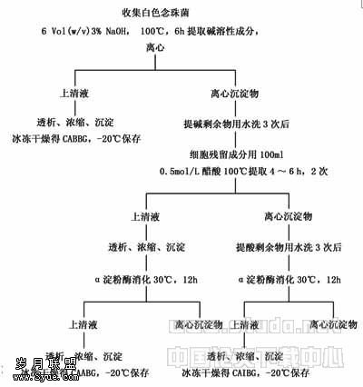
收集早期人胚胎51天标本3例,用4%多聚甲醛固定后制作8μm厚的石蜡切片。B-FGF抗体(Sigma公司产品),ABC药盒(Vector公司产品)。
1.2 方法
免疫细胞化学染色(immunocytochemistry staining ICC):间隔取石蜡切片,3例人胚胎标本取30张切片,入60℃烤箱0.5 h,梯度酒精脱水,常规H-E染色。抗原修复,ABC免疫组化染色。用0.3% H2O2预处理5 min,消除内源性过氧化物酶,用0.01 mol/L PBS漂洗3次,每次5 min。分别用兔抗B-FGF(效价1∶250,Sigma)抗体孵育切片,37℃ 1 h,入冰箱(4℃)过夜。用0.01 mol/L PBS漂洗3次,每次5 min。用生物素标记的羊抗兔血清(1∶200 Vector公司产品)孵育,37℃ 2 h。用0.01 mol/L PBS漂洗3次,每次5 min。用卵白素-生物素复合物(1∶100 Vector公司产品)孵育,37℃ 1 h。0.05 mol/L TBS漂洗3次,每次5 min。切片入DAB-H2O2显色液,室温反应5 min。(显色液配制:用0.05 mol/L TBS 20 ml溶解DAB 10 mg,充分摇匀,使DAB终浓度为0.05%,显色前加入30%的H2O2 8μl,使其终浓度为0.01%)。0.01 mol/L PBS漂洗3次,每次5 min。梯度酒精逐级脱水,二甲苯透明,树胶封片。
1.3 对照实验
取正常兔血清代替兔抗B-FGF血清。其它步骤与上述相同。
2 结果
2.1 肝
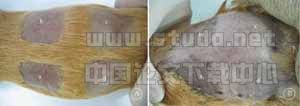
胚胎肝细胞呈不规则团索状,B-FGF阳性细胞呈点状和团块状散在分布于整个肝组织内,以后者居多。阳性细胞大多为肝细胞,部分为造血细胞,阳性反应聚集成团 (见图1)。
2.2 表皮
在胚胎表皮可见B-FGF阳性反应物,其细胞反应强弱和数目在胚胎的不同部位变化较大(见图2)。
2.3 上下肢芽
在胚胎上下肢芽的间充质中可见B-FGF阳性反应物(见图3)。
3 讨论
胚胎发育是一个十分复杂的过程,其过程受多种因子的调控,大量研究表明B-FGF作为一种营养因子,在胚胎发育的多个阶段都有重要作用,能诱导组织细胞的分化与成熟,对胚胎发育具有十分重要的作用。
本实验观察到B-FGF在肝表达,B-FGF阳性细胞呈点状和团块状散在分布于整个肝组织内。阳性细胞大多为肝细胞,部分为造血细胞,阳性反应聚集成团,提示B-FGF对肝细胞具有促进增殖的作用,同时可能具有旁分泌的作用。胚胎时期人的肝组织是合成B-FGF的主要部位,约占血液B-FGF总量的90%,肝细胞中B-FGF的表达,提示肝产生的大部分B-FGF分泌入血,对其他组织行使类似激素样内分泌作用。
B-FGF在表皮中表达,其细胞免疫阳性反应强弱在胚胎的不同部位变化较大,提示B-FGF对表皮细胞的增殖具有促进作用,胚胎发育的时序可能是在胚胎的不同部位免疫阳性反应强弱的原因。B-FGF的表达与胚胎上下肢芽发育过程有关。Martinou等[3]使用免疫组化及原位杂交方法在3-15天的鸡胚胎中观测到B-FGF及其mRNA在中胚层中表达最为明显。本实验观察到B-FGF在上下肢芽中表达,其间充质中可见B-FGF阳性反应细胞和纤维,提示B-FGF对上下肢芽的发育具有促进作用。
【】
[1]Christensen R N,Weinstein M,Tassava R A.Fibroblast Growth Factors in Regenerating Limbs of Ambystoma:Cloning and Semi-quantitative RT-PCR Expression Studies[J].Exp Zool,2001,290(5):529-540.
[2]Bates B,Hardin J,Zhan X,et al.Biosynthesis of Human Fibroblast Growth Factor-5[J].Mol Cell Biol,1991,11(4):1 840-1 845.
[3]Munoz-Sanjuan I,Simandl B K,Fallon J F,et al.Expression of Chicken Fibroblast Growth Factor Homologous Factor(FHF)-1 and of Differentially Spliced Isoforms of FHF-2 during Development and Involvement of FHF-2 in Chicken Limb Development[J].Development,1999,126(2):409-421.
[4]Gospodarrowicz D,Ferrara N,Schweigerer L,et al.Structural Characterization and Biological Functions of Fibroblast Growth Factor[J].Endocrine Rev,1987,8:95-104.
[5]National Cancer Institute,Cancer Genome Anatomy Project (CGAP),Tumor Gene Index http://www.ncbi.nlm.nih.gov/ncicgap.Contact: Robert Strausberg,Ph.D.Tel: (301) 496-1 550.
[6]Aoyagi A,Nishikawa K,and Saito H,et al.Characterization of Basic Fibroblast Growth Factor- mediated Acceleration of Axonal Branching in Cultured Rat Hippocampal Neuons[J].Brain Res,1994,661:117-126.
[7]Raz V,Kelman Z,Avivi A,et al.PCR-based Identification of New Receptors:Molecular Cloning of a Receptor for Fibroblast Growth Factors[J].Oncogene,1991,6:753-760.
[8]Yayon A,Zimmer Y,Hong SG,et al.A Confined Variable Region Confers Ligand Specificity to Fibroblast Growth Factor Receptors[J].EMB,1992,11:1 885-1 890.